Printable Canine Brain Diagram Here we present a canine brain atlas derived as the diffeomorphic average of a population of fifteen mesaticephalic dogs The atlas includes 1 A brain template derived from in vivo T1 weighted imaging at 1 mm isotropic resolution at 3 Tesla with and without the soft tissues of the head 2 A co registered high resolution 0 33 mm
This anatomical module of vet Anatomy is based on sections of the normal canine brain on MRI More than 400 images are available They have been obtained on a 2 years old healthy labrador retriever on a 1 5 T MRI Images are organized as 22 spin echo transverse section images with 5 weightings available FLAIR T1 T1w Gd T2 T2 w Canine Brain MRI Brain Tissue Atlas presents transverse views of a Beagle Brain obtained by Magnetic Resonance Imaging To facilitate neuroanatomy identification T2 weighted T1 weighted and Proton Density MRIs are paired with stained tissue sections obtained from a different dog brain
Printable Canine Brain Diagram
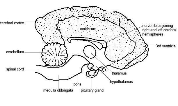 Printable Canine Brain Diagram
https://www.hannegrice.com/wp-content/uploads/Anatomy_and_physiology_LS_dogs_brain.jpg
In this manuscript we create and make available a detailed MRI based cortical atlas for the canine brain
Pre-crafted templates provide a time-saving solution for producing a varied variety of files and files. These pre-designed formats and designs can be made use of for various personal and professional jobs, consisting of resumes, invites, leaflets, newsletters, reports, discussions, and more, enhancing the content development procedure.
Printable Canine Brain Diagram

Printable Puppy Teeth Chart Printable World Holiday
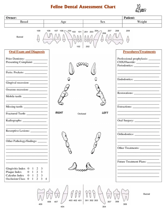
Printable Canine Dental Chart
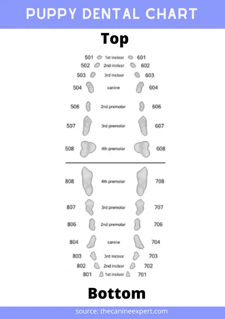
Printable Puppy Teeth Chart Customize And Print
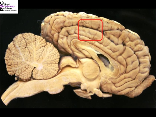
Canine Brain Dissection Anatomy Resource WikiVet English

Canine Dental Chart Printable Printable World Holiday

Canine Brain Anatomy Movement Science Motor Control At Queen Mary

https://anatomylearner.com/dog-nervous-system
25 04 202316 07 2022by anatomylearner The dog nervous system is divided into the central and peripheral nervous systems The brain and the spinal cord include the central nervous system CNS In contrast the canine peripheral nervous system PNS consists of cranial nerves spinal nerves and visceral peripheral components
https://www.merckvetmanual.com/dog-owners/brain,-spinal-cord,-and
The brain is divided into 3 main sections the brain stem which controls many basic life functions the cerebrum which is the center of conscious decision making and the cerebellum which is involved in movement and motor control

http://vanat.cvm.umn.edu/WebSitesNeuro.html
This web app presents a stand alone interactive tutorial for canine cranial nerves and cranial nerve nuclei including a cranial nerve innervation summary table canine brain attachment sites of cranial nerve roots identification of nuclear neuron columns per fiber type in brainstem cartoons cranial nerve nuclei shown within brainstem
http://vanat.cvm.umn.edu/brainsect
Two modes are featured 1 focus on names and see the components labeled and described or 2 focus on components and see the names high lighted An index screen showing small images of all sections is provided for navigation to particular transection images Note Relative magnifications of the individual transection images is variable

https://resources.saylor.org/wwwresources/archived/site/wp-con…
The central nervous system CNS which consists of the brain and spinal cord The peripheral nervous system PNS which consists of the nerves that connect to the brain and spinal cord cranial and spinal nerves as well as the autonomic or involuntary nervous system Diagram 14 5 The nervous system of a horse
Full text Full text is available as a scanned copy of the original print version Get a printable copy PDF file of the complete article 4 8M or click on a page image below to browse page by page Click on the image to see a larger version The dog brain which can be used for neuroimaging studies MATERIALS AND METHODS Imaging was performed on a Philips Ingenia 3 0 T whole body MR machine Philips Medical Systems Best The Netherlands with a Philips SENSE Flex Medium coil using a 3D Turbo Field Echo sequence TR 9 85 ms
Brain template related label maps are essential for functional magnetic resonance imaging fMRI data analysis to localize neural responses In this study we present a detailed individual based T1 weighted MRI based brain label map used in dog neuroimaging analysis