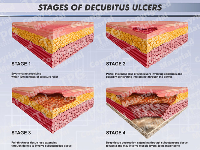
Pictures Of Stage 2 Decubitus Ulcers Printable Stage II A partial thickness wound presenting as a shallow open ulcer with a red pink wound bed May also present as an intact or open ruptured serum filled or serosanguinous filled blister Slough may be present but does not obscure the depth of
Bedsore Enlarge image Bedsores also called pressure ulcers and decubitus ulcers are injuries to skin and underlying tissue resulting from prolonged pressure on the skin Bedsores most often develop on skin that covers bony areas of the body such as the heels ankles hips and tailbone Decubitus ulcers also termed bedsores or pressure ulcers are skin and soft tissue injuries that form as a result of constant or prolonged pressure exerted on the skin These ulcers occur at bony areas of the body such as the ischium greater trochanter sacrum heel malleolus lateral than medial and occiput
Pictures Of Stage 2 Decubitus Ulcers Printable
 Pictures Of Stage 2 Decubitus Ulcers Printable
https://i.pinimg.com/736x/c4/9c/7f/c49c7f10c5eecd72a012f9287f56c7e5--deep-tissue-pressure-ulcer.jpg
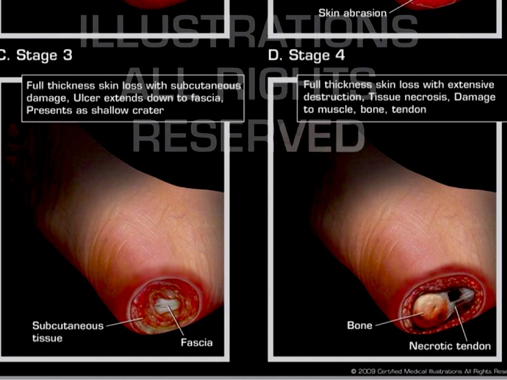
Decubitus ulcers occur in stages There s a staging process to help your healthcare professional diagnose and treat you Stage 1 and 2 ulcers usually do not require surgery but stage 3 and 4
Templates are pre-designed files or files that can be used for numerous purposes. They can save time and effort by supplying a ready-made format and design for producing different type of content. Templates can be used for personal or professional projects, such as resumes, invites, flyers, newsletters, reports, discussions, and more.
Pictures Of Stage 2 Decubitus Ulcers Printable

Leg Discoloration Stasis Dermatitis Health Life Media

Stages Of Decubitus Ulcers Order

Bedsores Also Called Pressure Ulcers Or Decubitus Ulcers Are Areas Of

Bed Sores Pressure Sores Doolan Platt Setareh LLPDoolan Platt

Stage 2 Pressure Ulcer Pictures Pictures Photos

Pressure Ulcer Classification

https://www.gettyimages.com/photos/decubitus-ulcer
Browse Getty Images premium collection of high quality authentic Decubitus Ulcer stock photos royalty free images and pictures Decubitus Ulcer stock photos are available in a variety of sizes and formats to fit your needs

https://www.clwk.ca/get-resource/pressure-ulcers-stages-with-photo…
Stage I ulcer will appear different than the colour of surrounding skin eschar Indicates the patient is at risk for further tissue damage if pressure is not relieved Partial thickness wound presenting as a shallow open ulcer with a red pink wound bed May also present as an intact or open ruptured serum filled filled blister

https://www.merckmanuals.com/home/multimedia/image/stage-2-pressure
Stage 2 Pressure Sore Buttocks This photo shows a stage 2 pressure sore on the upper right buttock arrow Tissues beneath the sore cannot be seen BOILERSHOT PHOTO SCIENCE PHOTO LIBRARY

https://www.steris.com//surgical-equipment/pressure-ulcer-stages-prevention
Stage 2 Pressure Ulcer Stages Stage II pressure ulcer involves partial thickness skin loss of the epidermis and may include the dermis The ulcer is superficial and looks like an abrasion blister or shallow crater Symptoms are pain swollen skin warmth and or red The sore may ooze clear fluid or pus

https://www.facs.org/media/32hdibi4/wound_pressure_ulcers.pdf
Your Pressure Ulcer Wound Home Skills Kit Pressure Ulcers Your Pressure Ulcer 8 Stage 2 Signs The skin is broken The ulcer is pink or red at the center The skin may be shiny dry or have a blister that is draining What to do Stay off the area and remove all pressure Contact your health care provider right away
Category Stage 2 Partial thickness Partial thickness loss of dermis presenting as a shallow open ulcer with a red pink wound bed without slough May also present as an intact or open ruptured serum filled or sero sanginous filled blister Presents as a shiny or dry shallow ulcer without slough or bruising This Clinical Image section of this site is a visual educational resource dedicated to providing pictures that are representative of common and uncommon physical exam findings Discussions of pathophysiology diagnostics and treatment are not included Content has been curated by Dr Goldberg and Staff
Stage 2 A shallow wound with a pink or red base develops You may see skin loss abrasions and blisters Stage 3 A noticeable wound may go into your skin s fatty layer the hypodermis Stage 4 The wound penetrates all three layers of skin exposing muscles tendons and bones in your musculoskeletal system What are the complications